SAR405 from Selleck Target VPS34 Autophagy Inhibition Activates STING NF-kB Pathway
Copy code to add this image to your page
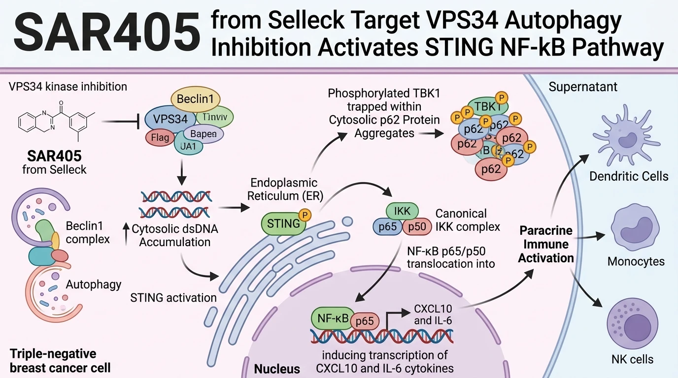
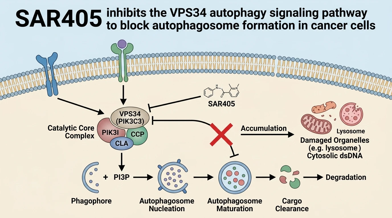
Targeting autophagy in malignancies with high baseline autophagic flux reveals key nodes linking organellar homeostasis to innate immune recognition. Pharmacological suppression of VPS34 (PIK3C3)—the catalytic subunit of the Class III phosphatidylinositol 3-kinase complex responsible for generating phosphatidylinositol 3-phosphate (PI3P)—disrupts early endosomal sorting and autophagosomal nucleation. In high-autophagy triple-negative breast cancer (TNBC) cellular models, applying the selective kinase inhibitor SAR405 blocks autophagic flux. This metabolic block prevents the turnover of damaged organelles and impedes the clearing of cytosolic double-stranded DNA (dsDNA). Under baseline conditions, cytosolic dsDNA is degraded via autophagosomal sequestration. Inhibiting VPS34 causes intracellular accumulation of dsDNA fragments within the cytoplasm.
Accumulated cytosolic dsDNA engages cyclic GMP-AMP synthase (cGAS), generating the second messenger cGAMP to activate the stimulator of interferon genes (STING) protein on the endoplasmic reticulum membrane. Classical STING activation recruits TANK-binding kinase 1 (TBK1) to phosphorylate interferon regulatory factor 3 (IRF3), triggering type I interferon (IFN-I) transcription. However, cellular context alters this downstream cascade under selective VPS34 inhibition. Structural characterization of the VPS34 complex kinase domain provides detailed insight into how lipid kinase inhibition disrupts intracellular traffic. In SAR405-treated TNBC cells, activated TBK1 (pTBK1) undergoes spatial redistribution rather than free cytoplasmic propagation. Autophagy failure leads to accumulation of p62/SQSTM1 sequestosome aggregates. These cytoplasmic p62 bodies physically trap phosphorylated TBK1.
Sequestration of pTBK1 in p62 aggregates disconnects STING from the canonical IRF3-mediated IFN-I branch. The physical compartmentalization of pTBK1 prevents IRF3 phosphorylation and nuclear translocation. Despite this suppression of IFN-I signaling, STING engagement continues to drive robust immunostimulatory outputs. The pathway redirects through a TBK1-independent canonical NF-κB signaling sequence. STING activation recruits the IκB kinase (IKK) complex, promoting IκBα phosphorylation and degradation. Free p65/p50 heterodimers translocate into the nucleus to drive transcriptional programs.
Quantitative gene expression assays demonstrate marked induction of pro-inflammatory cytokines and chemokines, specifically CXCL10 and IL-6. This chemokine upregulation occurs independently of type I interferon feedback loops. Importantly, while classical IFN-I signaling upregulates programmed death-ligand 1 (PD-L1), the SAR405-induced NF-κB pathway does not alter basal PD-L1 expression levels. The stability of immune checkpoint expression prevents premature T-cell exhaustion pathways. The resulting secretome from SAR405-treated TNBC cell lines activates innate immune effectors. In vitro conditioned media assays show robust functional activation of primary human monocytes, dendritic cells, and natural killer (NK) cells. Pharmacological VPS34 inhibition produces stronger immune cell activation than ionizing radiation alone under tested experimental conditions.
Selective Signaling Re-routing and Innate Immune Activation Dynamics
Copy code to add this image to your page
SAR405 selectively inhibits the Class III PI3K VPS34, suppressing autophagic degradation and causing cytosolic dsDNA accumulation in high-autophagy TNBC cells. This activates cGAS-STING signaling. Rather than inducing canonical IRF3-mediated type I interferon transcription, active pTBK1 is sequestered within cytosolic p62 aggregates. Signaling redirects through a TBK1-independent canonical IKK/NF-κB axis. This molecular switch drives strong expression of immunostimulatory chemokines, such as CXCL10 and IL-6, without upregulating the immune checkpoint PD-L1, ultimately boosting innate immune cell activation.
Sequestration of Phosphorylated TBK1 Within p62 Aggregates
Inhibition of VPS34 by SAR405 blocks autophagic degradation pathways. Undegraded cargo accumulates rapidly. Cytoplasmic p62/SQSTM1 forms dense protein aggregates throughout the cytosol. Phosphorylated TBK1 binds these p62 clusters, remaining physically trapped within the insoluble protein fraction. In vitro kinase assays confirm that TBK1 maintains enzymatic activity; however, localized compartmentalization prevents interaction with soluble IRF3 substrates. Consequently, classical type I interferon transcription fails to initiate. This spatial isolation effectively disconnects STING from the canonical IFN-β induction pathway in TNBC models.
STING Activation Induces Pro-Inflammatory Chemokines Without PD-L1 Upregulation
Cytosolic dsDNA persistence activates the cGAS-STING axis continuously. Applying SAR405 from Selleck drives signaling through an alternative canonical IKK-NF-κB cascade. Phosphorylation of IκBα proceeds normally, allowing nuclear entry of the p65 subunit. Transcriptional profiling reveals sharp elevations in CXCL10 and IL-6 mRNA levels. High-affinity CXCL10 expression establishes chemotactic gradients. Notably, cell surface expression of PD-L1 remains completely unchanged during this inflammatory conversion. The absence of checkpoint upregulation preserves effector cell responsiveness within the local microenvironment.
Immune Activation in Triple-Negative Breast Cancer Supernatants
Conditioned media from TNBC cells treated with SAR405 from Selleck exhibits potent immunostimulatory properties. Primary human monocytes exposed to these supernatants undergo rapid phenotypic differentiation into activated antigen-presenting phenotypes. Dendritic cells demonstrate elevated co-stimulatory marker expression. Natural killer cell cytotoxicity assays show increased granzyme B secretion. Comparative experiments confirm that pharmacological autophagy blockade induces higher levels of innate immune stimulation than single-dose focal radiation.