Volasertib PLK inhibitor BI6727 attenuates pulmonary vascular remodeling via target PLK1 suppression
Copy code to add this image to your page
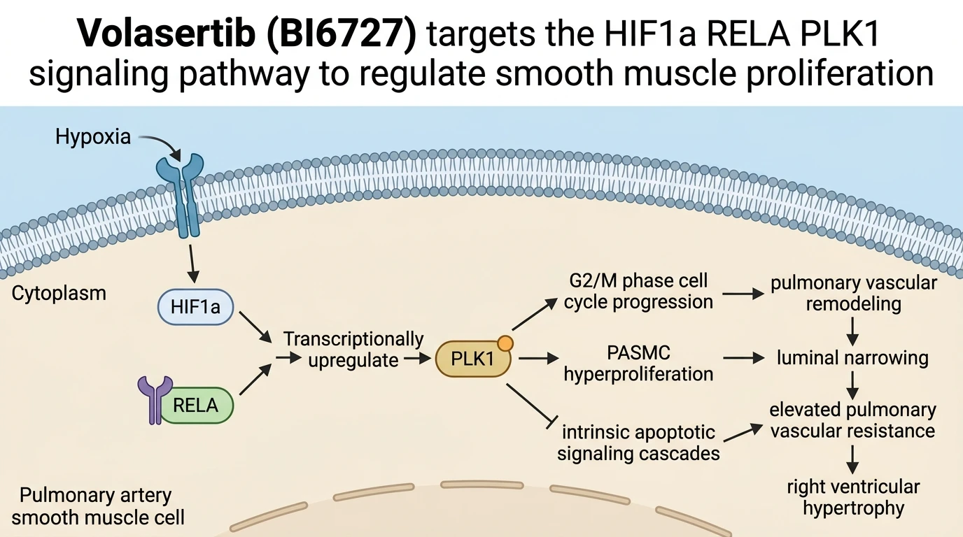
Pulmonary arterial hypertension (PAH) is characterized by progressive structural remodeling of small pulmonary arteries, leading to sustained increases in pulmonary vascular resistance and right ventricular failure. At the cellular level, hyperproliferation and apoptosis resistance of pulmonary artery smooth muscle cells (PASMCs) constitute primary drivers of vascular lumen narrowing. Recent mechanistic investigations identify Polo-like kinase 1 (PLK1)—a serine/threonine kinase recognized for regulating mitotic entry, centrosome maturation, and spindle assembly—as a crucial pathological node in pulmonary vascular remodeling.
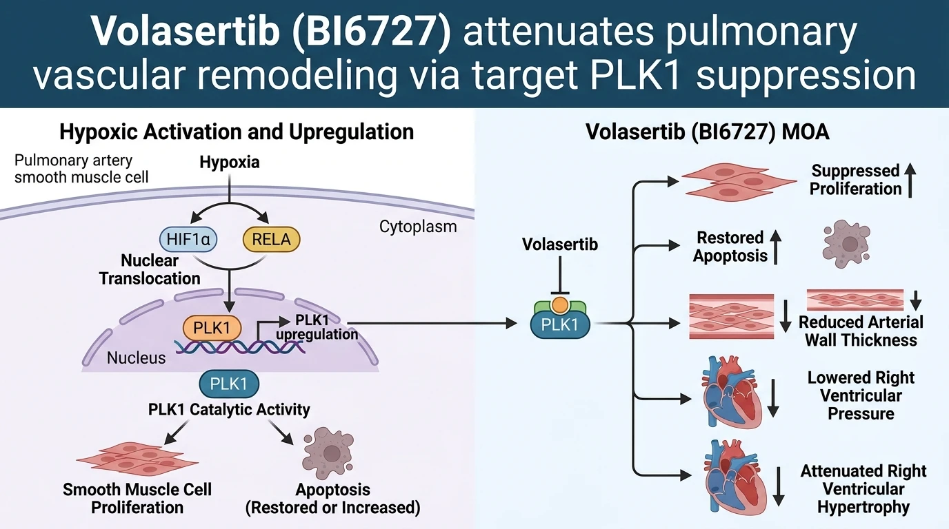
Under normoxic conditions, PLK1 expression remains low in non-cycling vascular tissue. However, chronic hypoxia triggers a distinct transcriptional cascade that markedly upregulates PLK1 in PASMCs. Exposure to low ambient oxygen tension stabilizes the alpha subunit of hypoxia-inducible factor 1 (HIF1a). Simultaneously, hypoxic stress activates nuclear factor kappa B signaling, mobilizing the RELA (p65) subunit. Functional promoter analyses demonstrate that HIF1a and RELA co-occupy specific response elements within the PLK1 promoter region, acting synergistically to drive high-level PLK1 transcription. This hypoxia-activated HIF1a/RELA signaling axis directly links environmental oxygen sensing to cell cycle regulatory machinery in vascular smooth muscle.
Targeted inhibition of PLK1 offers a novel mechanism to disrupt PASMC accumulation. The small-molecule compound Volasertib (BI6727), a potent ATP-competitive PLK1 inhibitor, selectively binds the catalytic domain of PLK1, preventing downstream phosphorylation events necessary for mitotic progression. In cellular assays using primary human PASMCs cultured under hypoxic conditions, target engagement by the Volasertib PLK inhibitor reinstates pro-apoptotic signaling. Inhibiting PLK1 activity leads to mitotic arrest, activation of caspase-3, and induction of apoptotic death specifically in rapidly dividing smooth muscle populations. Conversely, non-proliferating vascular cells maintain viability at equivalent compound concentrations, establishing a therapeutic window in vitro.
The efficacy of Volasertib (BI6727) has been rigorously examined in vivo using the Sugen5416/hypoxia rodent model of severe pulmonary hypertension. Administration of BI 6727 effectively attenuates disease progression across multiple physiological and histological metrics. In treated animals, right ventricular systolic pressure (RVSP)—a surrogate marker for pulmonary arterial pressure—decreases significantly compared to vehicle-treated controls. This reduction in pressure corresponds with reduced right ventricular strain, as evidenced by a lower Fulton index (right ventricle to left ventricle plus septum mass ratio, RV/LV+S).
Histological evaluation of lung sections reveals that PLK1 inhibition limits vascular remodeling within resistance arteries. Treatment with BI 6727 reduces medial arterial wall thickness and decreases the proportion of fully muscularized distal arterioles. Beyond structural vessel changes, PLK1 inhibition dampens inflammatory and fibrotic gene expression profiles within both pulmonary and cardiac tissues. Transcription levels of key pro-inflammatory cytokines, including interleukin-6 and tumor necrosis factor-alpha, drop following drug administration. Similarly, markers of collagen deposition and extracellular matrix remodeling are downregulated in the right ventricle, attenuating myocardial fibrosis.
Hemodynamic parameters demonstrate secondary benefits on cardiac function. Echocardiographic measurements indicate that Volasertib (BI6727) treatment improves right ventricular stroke volume, cardiac output, and ejection fraction in Sugen5416/hypoxia-exposed mice. By reducing pulmonary vascular impedance, PLK1 inhibition alleviates right ventricular afterload, preventing compensatory hypertrophy from transitioning into overt right heart failure.
While these preclinical observations highlight the therapeutic viability of target PLK1 suppression, several boundary conditions warrant consideration. The pharmacokinetic and safety profiles of Volasertib (BI6727) have been primarily characterized in oncological settings, where intermittent high-dose schedules are common. Adapting PLK1 inhibitor regimens for chronic cardiovascular indications like pulmonary hypertension necessitates evaluating low-dose continuous dosing strategies to minimize potential myelosuppression. Furthermore, while the Sugen5416/hypoxia murine model replicates key features of severe human pulmonary hypertension, findings must ultimately be validated in human tissue explants and clinical trial cohorts. Nevertheless, current empirical evidence demonstrates that targeting the HIF1a/RELA/PLK1 axis represents a novel approach to reversing vascular remodeling and preserving right ventricular function.
Hemodynamic and Histological Amelioration in Murine Models
Copy code to add this image to your page
Polo-like kinase 1 (PLK1) overexpression, mediated by hypoxic activation of the HIF1a/RELA complex, drives pulmonary artery smooth muscle cell hyperproliferation and apoptosis resistance. Administration of the small-molecule inhibitor Volasertib (BI6727) selectively inhibits PLK1 kinase activity. This molecular engagement blocks downstream mitotic signaling, triggers apoptosis in proliferating vascular smooth muscle cells, reduces pulmonary arterial wall thickness, and lowers right ventricular systolic pressure in experimental rodent models of pulmonary hypertension.
HIF1a and RELA Transactivate PLK1 Under Hypoxia
Hypoxia serves as a primary driver of pulmonary vascular pathology. Chronic oxygen deprivation stabilizes hypoxia-inducible factor 1-alpha (HIF1a), which subsequently forms a functional transcriptional complex with the RELA subunit of nuclear factor kappa B (NF-kB). Within human pulmonary artery smooth muscle cells, this complex binds directly to promoter elements of the Polo-like kinase 1 gene. Elevated kinase expression accelerating mitotic entry follows. Increased cellular density reduces susceptibility to apoptotic stimuli. The small-molecule agent Volasertib PLK inhibitor prevents kinase-domain transphosphorylation in vitro. In cultured primary PASMCs, target inhibition halts proliferative expansion.
Attenuation of Right Ventricular Pressure in Sugen5416 Mice
In vivo efficacy was evaluated in Sugen5416/hypoxia-exposed mice exhibiting established pulmonary hypertension. Oral dosing of Volasertib (BI6727) altered disease progression markedly over the treatment period. Right ventricular systolic pressure dropped significantly compared to vehicle controls. Morphometric analysis demonstrated a concurrent reduction in the right ventricular hypertrophy ratio (RV/LV+S). Medial wall thickening within distal pulmonary arteries diminished. Short term target engagement suppressed local inflammatory cytokines. Furthermore, cardiac output and ejection fraction exhibited measurable recovery. These findings confirm functional reverse remodeling within the pulmonary vasculature.